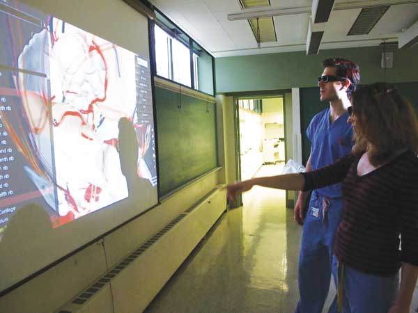
BY TERENCE CONFINO | In the constantly evolving field of medical science, N.Y.U. School of Medicine has stepped up its curriculum by combining science with 3D computer technologies.
Dr. Sally Frenkel is just one associate professor that worked in partnership with BioDigital Systems LLC to create the BioDigital Human, a 3D interactive virtual cadaver that aids medical students’ understanding of dissection and anatomy.
“The first thing to say is that nothing actually compares with the experience of dissecting an actual cadaver,” said Frenkel. “But the 3D technology comes close.”
Students are using consumer-grade 3D glasses to view life-size digital content displayed on a projector screen in the N.Y.U. anatomy lab. They can also use laboratory iPads to magnify and explore human anatomy in a way that the more static two-dimensional sources, like textbooks and dissection manuals, simply can’t.
The new teaching relies heavily on simulation, and one of the advantages the 3D technology provides is spatial awareness.
With the virtual cadaver, students can zoom in on any organ and spin it to view it from their angle of choice. They can also reveal and hide layers of muscle, bone and nerves and then use tools to dissect or analyze them.
“It allows you to manipulate and rotate the body organ whatever way you like and you can dissect in layers. First the skin, then muscles, then blood vessels,” said Frenkel, who has been with N.Y.U.’s School of Medicine for nearly 20 years.
The use of lab iPads “allows for mastery of the subject matter,” said Frenkel, since students can continually review any area they feel weak in as much as they like.
Victoria Fang, 23, is a first year M.D. / Ph.D. candidate at N.Y.U. School of Medicine. She is interested in pursuing a career in oncology research and medicine, the branch of medical science that deals with tumors, including their origin, development, diagnosis and treatment.
“When I first heard about [the 3D virtual cadaver] in late August, it was still in the works and it sounded amazing,” Fang said.
She went on to note the obvious disadvantages to using old books, as she put it, “following line by line, when you’re not sure of something, then having to go to two to three other books to get the information.” As opposed to looking at illustrations and photos in books, it’s actually much easier to just click on an image and zoom in to see it, said Fang.
“The books have more difficult spatial concepts, things become difficult to visualize in that sense, and the 3D technology is incredibly helpful,” she noted.
When asked about her first impressions when confronted with the School of Medicine’s high-tech approach to science, Fang said, “It was amazing in terms of what technology can do.”
The technological upgrade comes as part of N.Y.U. School of Medicine’s new curriculum for the 21st century or “C21.” These changes are the result of dramatic advances in the healthcare delivery system, which have, in turn, prompted curricular reforms in medical education.
Dr. Frenkel hopes that the new digital environment will result in the reduction of medical errors and improved patient care.
She likened her students’ technological experience to a future where the doctor stands by a patient’s bedside using an iPad to explain treatment options.
However, some things will just never change. And as both professor and student agree, nothing comes close to dissecting an actual human cadaver.
“Absolutely nothing can really replace it,” Frenkel said. “The cadaver provides an intimate experience in providing students with the feel and relationship of the human body. We consider it the students’ first patient.”
Medical student Fang concurred.
“To be honest, the dissection itself is what allows you to learn the actual material,” she said. “A computer display is absolutely not what we’re encountering with patients.
“And as far as experience goes,” Fang concluded, “unless you go into surgery — it’s probably the only time we’ll get to see the inside of the body.”


